

 19. August 2026
 Boosting FLIM for Next-Generation Cancer Diagnostics
Cancers developing in epithelial tissues lining account for a large share of new cancer cases worldwide – and consequently to cancer-related deaths. (1) Early and accurate detection by endoscopy or biopsy is essential for successful treatment. However, current diagnostic methods rely mostly on visual inspection and standard white light, combined with contrast agents, fluorescence, dyes, and molecular probes. Those approaches are limited to detecting only specific targets and come with complex workflows, regulatory burdens, and long procedure times. This makes accurate diagnosis difficult if a tumor does not express the expected marker or several pathological changes coexist.
Identification of multiple pathological states in a single examination without markers or contrast agents would be significant step forward for cancer diagnostics. Marker-free optical technologies promise this capability, but they are not clinically viable yet. To close this gap, it is necessary to advance tissue scanning technologies for faster, broader, and more informative data collection without increasing procedure time or complexity.
The Challenges
Accurate diagnosis requires information-rich datasets that capture spatial and temporal variations across tissue for reliable analysis. Endoscopy and biopsy procedures demand fast data acquisition and analysis, as the time available for the examination is limited because of patient safety and tissue viability. Most current time-resolved imaging approaches, such as point- or line-scanning FLIM systems, achieve high spatial and temporal resolution by acquiring data sequentially. As a result, it is not practical to scan larger tissue areas with point-level resolution to generate large datasets with reliable lifetime information within clinically viable time periods.
Consequently, the following three challenges need to be addressed:
#1 Expand the scanned tissue area
#2 Analyze the information from that area in point-by-point resolution
#3 Pre-process and compress the data close to the detector to overcome limitations in data bandwidth and latency
Our Solution: Boosted FLIM
Fluorescence Lifetime Imaging Microscopy (FLIM) is a powerful tool for tissue diagnostics and has been in use for more than 20 years. FLIM combines fluorescence lifetime spectroscopy with microscopy, allowing researchers to visualize how long fluorophores stay in their excited state across different regions of a sample. Instead of measuring how bright a signal is (fluorescence intensity), FLIM measures how long fluorescence lasts (decay time/fluorescence lifetime), providing contrast based on molecular environment rather than fluorophore concentration.
Typically, FLIM systems use a pulsed laser in the femto- or picosecond range as an excitation source together with time-domain methods such as time-correlated single-photon counting (TCSPC), time-gating or pulse sampling, or frequency-domain techniques, depending on the application and required speed, to measure lifetimes.
Picosecond lasers are used for one-photon excitation of standard fluorophores. As the intrinsic fluorescence is 100ps -few ns, the pulse width must be short enough, e.g. in the range of 10-100ps. Picosecond lasers are generally easier to implement due to their smaller footprint and less safety concerns – and they are cheaper.
Femtosecond lasers are mostly used for two-photon or multiphoton FLIM because those methods require very high peak intensities for the nonlinear absorption, and femtosecond lasers allow for the necessary peak power while keeping the average power low. They are also suitable for deep and thick tissue imaging.
A confocal microscope is then used to collect spatially resolved data. However, as conventional FLIM setups rely on a single laser source and a single-channel time-resolved detector, such as photomultiplier tubes (PMTs), data acquisition is inherently limited to point-by-point or line-by-line scanning.
Covering larger areas of tissue with this approach would take more time than is feasible for examining live patients or fresh tissue samples. This sequential acquisition fundamentally limits throughput and prevents real-time or near–real-time FLIM in clinical environments.
How It Works
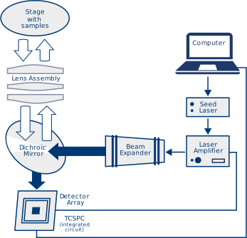
Our boosted FLIM approach overcomes the limitations of conventional FLIM by collecting more data per unit time and analyzing larger tissue areas in parallel, both spatially and spectrally. The central idea is to replace point-scanning detection with wide-field, parallel acquisition using a two-dimensional SPAD (Single-Photon Avalanche Diode) array. This allows fluorescence lifetime acquisition across an extended field of view simultaneously rather than sequentially pixel-by-pixel.
By imaging a much larger tissue area, the system dramatically reduces acquisition time while maintaining the temporal resolution required for accurate lifetime estimation. In combination with high-speed readout and timing electronics, the architecture supports rapid TCSPC or time-gated detection at each pixel, enabling near–real-time FLIM.
Using picosecond excitation (pulse durations on the order of 50–100 ps) with narrowband spectral resolution (~0.5 nm per channel/pixel), the system supports wide-field fluorescence lifetime acquisition at repetition rates in the tens of megahertz. Image fields of views on the order of several hundred micrometers can be achieved using detector arrays with hundreds of pixels per dimension, with per-pixel integration times ranging from sub-millisecond to a few seconds depending on the fluorescence photon budget of the sample. Importantly, the system operates at average laser powers in the low- to mid-tens of milliwatts, sufficient for reliable lifetime estimation while remaining within established photodamage and safety limits. The marker-free method leverages the intrinsic autofluorescence of tissue cells. It captures both the full fluorescence spectrum excited by the laser wavelength and the corresponding decay times across the field of view, enabling classical marker-based diagnostics as well as marker-free detection of multiple tumor types in a single scan.
On the laser side, we use a neoLASE MOPA laser system that delivers a wider beam to cover larger tissue areas at once instead of scanning individual points. The increased illumination area and higher photon flux improve signal-to-noise ratio and allow reliable lifetime estimation at significantly shorter acquisition times, while remaining within photodamage and safety limits.

On the detector side, the imaging device simultaneously acquires data from many pixels. To achieve this, we use a SPAD array from our partner Singular Photonics that enables ultra-fast, high-sensitivity photon detection in a compact format. Each pixel performs lifetime acquisition in parallel, removing the bottleneck of sequential scanning.
Crucially, photon timing data is processed and compressed directly on the detector chip. This means the limited data transfer rate to the FPGA is no longer a system constraint, enabling high-throughput FLIM suitable for dynamic biological processes and time-critical diagnostic workflows.
The combination of an amplified laser and a 2D SPAD array supports near-real-time processing, parallel spatial and spectral analysis, and high throughput acquisition, making FLIM faster, more comprehensive, and better suited for clinical applications than ever before.
SPAD Technology: The Core Advantage
Single Photon Avalanche Diodes (SPADs) are very sensitive light detectors capable of registering individual photons. They can detect extremely low levels of light with very high timing precision, usually in the range of tens of picoseconds. This makes them an ideal match for applications with high requirements for sensitivity, timing accuracy, and speed, such as time-resolved spectroscopy, FLIM, and quantum optics.
SPADs operate in Geiger mode above breakdown voltage. A single photon triggers an electron-hole pair, creating an avalanche that generates a digital pulse representing a detected photon event.
A quenching circuit stops the avalanche and resets the device after each event, followed by a short dead time until the system is ready for the next cycle.
When grouped into arrays, SPADs enable spatially resolved, parallel detection. Multiple SPADs can be combined into macro-pixels, improving photon detection efficiency and signal-to-noise ratio. They allow spatial binning and flexible resolution trade-offs, reduce the impact of dead time by distributing photon events, and support parallel readout for faster data acquisition. For example, the Andarta SPAD array from Singular Photonics is set up in a 4 × 4 pattern.

Modes of Operation
Our system supports multiple modes to meet different research and clinical needs. Histogram mode for FLIM includes on-chip data compression and histogram creation. Other available modes include photon counting, TCSPC, autocorrelation, time-gated, and burst mode. These modes can be switched, offering flexibility for diverse research and clinical applications.
SPAD Cameras and Modes of Operations
Our boosted FLIM system can be configured with two different SPAD cameras, each offering distinct acquisition modes to suit a range of applications:
Andarta SPAD array
This is a 512x 512 (128 x 128 binned) SPAD array designed for wide-field and general-purpose imaging. It supports time-gated acquisition, autocorrelation (pixel-level or ensemble), and burst mode. These modes make the Andarta array particularly suited for imaging dynamic processes including widefield fluorescence lifetime imaging, diffuse correlation spectroscopy (DCS) for blood flow measurements, rapid tissue imaging, and applications that require high temporal precision in transient or fluctuating signals. Further, we can combine time-gating and autocorrelation measurements, allowing integration of DCS-style blood flow analysis with traditional FLIM imaging or time-of-flight applications.

Sirona SPAD array
This is a 512 × 1 (16 blue SPADs per pixel, 16 red SPADs per pixel, selectable) SPAD array optimised for point- or line-scanning and spectrally resolved measurements. It supports TCSPC and histogram mode where photon arrival times are decoded and binned.
This makes the Sirona array ideal for applications needing high lifetime precision and robust delay analysis. This flexibility enables the cameras to address a wide range of research and clinical applications, from high-speed cancer imaging to cellular metabolism studies and dynamic physiological measurements.

Key Benefits
The boosted FLIM approach delivers more data per unit time compared to conventional FLIM. By combining wide-field acquisition with high temporal resolution, it enables marker-free diagnosis at clinically relevant speeds. Most importantly, it offers the chance to detect several tumor types in one examination.
We are actively seeking OEM partners and research institutes to bring our boosted FLIM technology to market. Together, we can revolutionize cancer diagnostics and make early detection faster, broader, and more accurate. Reach out to us and be part of the next big leap in medical imaging.
References
(1) More data on global cancer cases is available in the GLOBOCAN study from the World Health Organization (WHO): GCO Cancer Today
READ MORE ABOUT OUR ANALYTICS AND LASER SOLUTIONS

Discrete Detectors & Arrays
Browse Products >
Laser reliability, proven in the field
Read More >
LIBS and MOPA Lasers: More Efficiency for Aluminum Recycling
Read More >
Modular Laser Micromachining: A Scalable Platform for Precision Manufacturing
Read More >
Purity and Stability Meet High Power – 100 W Single-Frequency Laser
Read More >
Singular Photonics and AMS Technologies Join Forces to Bring Advanced SPAD Sensors to Europe
Read More >FAQ: Boosting FLIM for Cancer Detection
What is the main advantage of boosted FLIM compared to conventional FLIM?
Conventional FLIM systems typically acquire data point by point, which limits the area that can be examined within a practical time. Boosted FLIM combines a high-power laser source with a two-dimensional SPAD detector array to capture lifetime information across a much larger field of view simultaneously. This significantly reduces acquisition time while maintaining high temporal resolution.
Why are SPAD arrays important for next-generation FLIM systems?
SPAD (Single-Photon Avalanche Diode) arrays enable parallel detection of individual photons across many pixels at the same time. Combined with on-chip data processing, they support high-throughput fluorescence lifetime measurements without overwhelming downstream electronics. This makes real-time or near-real-time FLIM feasible for demanding research and diagnostic applications.
Can boosted FLIM support marker-free tissue diagnostics?
Yes. Boosted FLIM can use the natural autofluorescence of biological tissue instead of relying solely on fluorescent markers. By analyzing fluorescence lifetime and spectral information, the technology has the potential to distinguish different tissue types and support the detection of multiple tumor types in a single examination.
What role does the laser play in boosted FLIM?
The laser provides the excitation pulses required for fluorescence lifetime measurements. In boosted FLIM, a high-power MOPA laser system illuminates a larger tissue area than conventional point-scanning systems. The increased photon flux improves signal quality and enables reliable lifetime measurements with shorter acquisition times while remaining within established safety limits.
Is boosted FLIM available as a complete system?
AMS Technologies supplies key enabling technologies for OEMs and research organizations, including ultrafast laser systems, optical amplifiers, and SPAD detector technology. These components can be integrated into next-generation FLIM systems tailored to specific research or diagnostic applications.
Who should consider boosted FLIM technology?
Boosted FLIM is particularly relevant for OEMs developing advanced biomedical imaging systems, as well as research institutes working in fluorescence imaging, endoscopy, cancer diagnostics, and other life science applications where high-speed, high-resolution fluorescence lifetime measurements are required.
EXPLORE MORE
NEWS & EVENTS
AMS Technologies and neoLASE join B2Run Hannover
B2Run Hanover 2026 was fun! Thank you to EPIC Photonics, the organizing team – and most of all to our runners from AMS Technologies and ...
Read More >

From Lab Concept to Serial Production: Addressing the PIC Polishing Challenge with KrellTech NOVA
Why repeatable component loading is the first hurdle Every polishing process depends on how reliably a chip is loaded and secured. Repeatability in loading directly ...
Read More >

AIMEN Laser Week
October 26–29, 2026
O Porriño, ES
AIMEN Laser Week is an international event dedicated to laser technologies and their applications in industrial manufacturing, process monitoring, and analytical systems. The conference combines ...
Read More >
